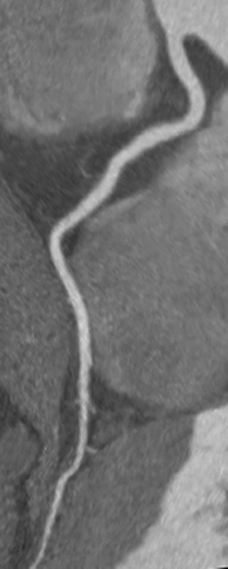
Researchers from the University of Erlangen in Germany and 22 other European centers aimed to show that high-iodine concentration contrast media are not superior to standard-concentration formulas, and they did so. The study randomized 452 patients from five countries to receive iobitridol 350 or one of two standard-concentration formulas. The average signal-to-noise ratio (SNR) and contrast-to-noise ratio (CNR) did not differ among the patient groups.
"The main finding is that minor differences in contrast agent concentration may not be all that important for image quality in coronary CTA," Achenbach toldAuntMinnieEurope.com. "No difference was found in regards to image quality, and importantly, diagnostic confidence," when comparing the three contrast agents.
The study is the largest to date to address the relationship between iodine concentration and image quality, the authors noted (European Radiology, 23 May 2016).
Contrast protocols are of key importance in CCTA, which is used to identify coronary artery stenosis as well as detecting and characterizing calcified and noncalcified plaque. Studies suggest lumen opacification of at least 300 HU is required to identify all components adequately, using a carefully administered contrast protocol that measures iodine delivery rate and provides enough iodine to do the job, but not too much.
Optimizing contrast
Several groups have looked at different CT attenuation levels with different contrast media protocols, but few have assessed the impact of iodine concentration on image quality, or determined whether differences of less than 50 mg/mL of iodine concentration could affect image quality.
Higher iodine concentrations produce greater opacification, but whether this benefits interpretation is unclear, especially considering that higher iodine concentrations raise potential risks for patients. So the optimal attenuation for CCTA is still being debated, the authors wrote.
Increasing total injected iodine "could raise safety issues for patients at risk such as contrast-induced nephropathy," they stated. "Therefore, adequate contrast enhancement must be balanced against the amount of iodine injected."
Three agents head to head
This study compared a contrast media formula with iodine concentration of 350 mg/iodine mL iobitridol (Xenetix, Guerbet) with two other contrast media formulations with higher iodine concentrations, including iopromide 370 mg/ml (Ultravist, Bayer Healthcare) and iomeprol 400 mg/ml (Iomeron, Bracco Diagnostics). The main objective was to demonstrate statistical noninferiority of the lower-concentration formula to the two alternatives.
The study examined 452 symptomatic adult patients (mean age 57.8) with suspected coronary artery disease who were scheduled for CCTA, in sinus rhythm, and without contraindications to beta-blocker medications, renal insufficiency, or previous coronary artery bypass grafts, stents, or artificial heart valves.
Patients received beta-blockers if their heart rate was above 65 bpm, and a minimum dose of 0.8 mg sublingual nitroglycerine spray before contrast medium injection and the scan. Contrast volume and delivery rate varied according to patient body weight from 4 ml/S for 60 kg and under; to 75 mL/S at 5 mL/S for 60 kg to 80 kg; and 90 mL at 6 mL/S for body weight greater than 80 kg. Injections were followed by a 100% saline flush.
Regions of interest of 2 mm2 were acquired in the left anterior descending (LAD) artery, the left circumflex (LCX) artery, and the left main coronary (LM) artery, as well as larger ROIs in the ascending aorta and left ventricle.
The two core lab readers reported stenosis on a five-point scale ranging from 5 (certainly yes) to 0 (certainly no) and the radiologists recommended patient management options including no action, medication, invasive coronary angiography, and other, the team wrote. Commercially available software (Comprehensive Cardiac Analysis, Philips Healthcare) was used to automatically track each coronary artery up to the distal segments.
Little difference
- The rate of fully evaluable scans was similar for all three agents, including 92.1% for iobritidol, 95.4% for iopromide, and 94.6% for iomeprol, the authors wrote.
- Iobitridol demonstrated its noninferiority over the best comparator agent with a 95% confidence index of the difference of [-8.8 to 2.1], with a prespecified noninferiority margin of -10%, the group wrote.
- Also, no difference was seen regarding the number of stenosis identified with the three contrast media agents. (p = 0.580).
- Multivariate analyses did show a relationship between calcium scoring and stenosis assessment according to territory (p < 0.001) and between calcium scoring and image quality regardless of territory (p = 0.007), the study team wrote.
- Average attenuation increased with higher iodine concentrations, but the average SNR and CNR did not differ between the three patient groups, Achenbach and colleagues wrote.
- The percentage of patients experiencing post CM-injection adverse events ranged from 15.1% for iobitridol, 19.5% for iopromide, and 15.1% for iomeprol. Most were cardiac disorders reported in EKG follow-up, the team wrote.
"This study demonstrated the noninferiority of iobitridol 350 in providing evaluable CT scans for assessment of coronary stenosis, as compared to CM with higher iodine concentrations" (iopromide 370 mg iodine/ml and iomeprol 400 mg iodine/ml).
The results also indicate iodine content can be reduced further, Achenbach et al wrote. As per common practice, iodine delivery rate was not adjusted, although doing so would have eliminated a key limitation.
 |
| Dr. Stephen Achenbach, University of Erlangen in Germany. Image courtesy of the European Society of Cardiology. |
The lack of a gold standard test such as invasive angiography means that diagnostic accuracy could not be compared among the three groups, but the limitation appears minor, according to Achenbach.
"While it would have been nice to have accuracy data, which means a comparison regarding the ability to detect stenoses, all parameters analyzed in this trial showed that the image quality and diagnostic confidence of the lower concentration contrast was not inferior," he wrote in an email. "It is hence rather safe to assume that diagnostic accuracy will also be equal."
The relatively low calcium burden in the population was another limitation. And injection rates were constant and not adjusted to achieve equal iodine delivery rates across the three groups.
"It was an additionally very comforting finding to see that in all patient groups within this study, the rate of 'fully evaluable' coronary CTA examinations was between 92% and 95%," Achenbach concluded. "Coronary CTA has become a very stable and robust imaging modality."
 |
| CCTA of a right coronary artery without stenosis or artifacts, imaged with iobitridol 350 mg iodine/mL. Image courtesy of Dr. Stephen Achenbach. |
No comments:
Post a Comment